


Medically reviewed by Jessica Churchill, MD | Reviewed June 2026
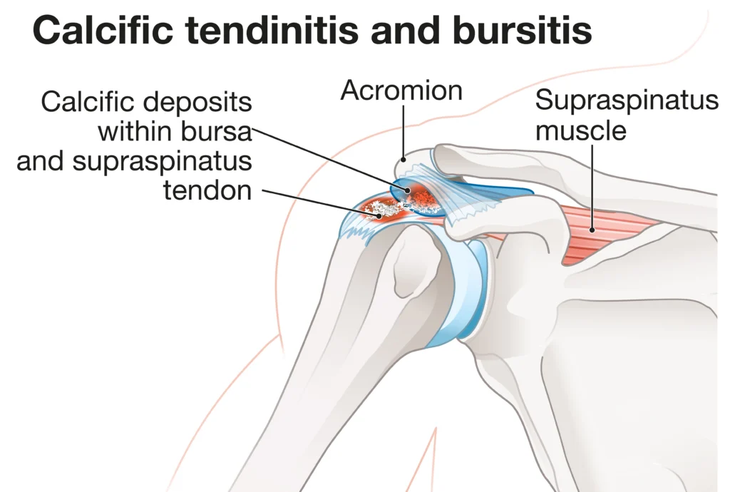
Shoulder pain that seems to come out of nowhere can be alarming. Patients often tell me they woke up one morning barely able to lift their arm, without any fall, accident, or obvious explanation. When I see that pattern in my clinic at Denver Health, calcific tendonitis is something I consider. Calcific tendonitis of the shoulder occurs when calcium deposits form within the tendons of the rotator cuff, most commonly in the supraspinatus tendon. Those deposits can cause significant inflammation, pressure, and pain. The good news is that many patients do not need surgery. Understanding what this condition is, why it happens, and what your options are can make a difference in how you navigate it.
Key Takeaways
- Calcific tendonitis of the shoulder involves calcium deposits in the rotator cuff tendons, most often the supraspinatus.
- Symptoms can range from a dull, chronic ache to sudden, severe pain, especially during the resorptive phase when the deposit breaks down.
- Non-surgical options, including physical therapy, anti-inflammatory medications, and ultrasound-guided needling, can be effective for many patients.
- Surgery is reserved for cases where conservative care has not provided adequate relief after a reasonable period of time.
What Actually Happens Inside the Tendon
The rotator cuff is made up of four muscles and their tendons that wrap around the shoulder joint and allow for lifting, rotating, and stabilizing the arm. In calcific tendonitis, calcium phosphate crystals accumulate within one or more of those tendons. The deposit typically forms in a region of the tendon with reduced blood supply.
The process tends to move through stages. During the formative phase, calcium builds up gradually. Many patients have no idea it is happening. The resting phase follows, when the deposit sits quietly. Then comes the resorptive phase, which is when the deposit begins to break down and the body tries to reabsorb the calcium. This phase can trigger an intense, acute inflammatory response. The calcium takes on a more fluid, toothpaste-like consistency, and the pressure inside the tendon rises sharply. This is when patients often show up in my office in significant pain.
After the resorptive phase, most deposits resolve and the tendon heals. That natural progression is part of why many patients improve without surgery. The challenge is that the resorptive phase can be unpredictable and may last weeks.
Who Tends to Develop This Condition
Calcific tendonitis tends to peak in adults between thirty and sixty years of age, and it appears more frequently in women than men, though it can occur in either.
Certain factors seem to increase the likelihood of developing calcific deposits. Repetitive overhead activity, whether from a job or a sport, can place sustained stress on the rotator cuff tendons. Thyroid disorders and metabolic conditions have also been associated with higher rates of calcific tendonitis, though the exact relationship is still being studied. In my experience, some of the patients I see with this condition have no obvious risk factors at all, which reinforces the point that this can happen to a wide range of people.
How Calcific Tendonitis Tends to Feel
The symptoms of calcific tendonitis of the shoulder can vary a lot depending on which phase the deposit is in. During the formative phase, many patients feel nothing or perhaps a vague aching that they attribute to a pulled muscle or general wear and tear. The pain may be mild and intermittent, and it often does not interfere much with daily activity.
When the deposit enters the resorptive phase, the picture changes. The pain can become severe and come on quickly. Lifting the arm, reaching overhead, and even lying on the affected shoulder at night can become difficult or impossible. Some patients describe the pain as one of the worst they have experienced.
A few symptoms that commonly accompany calcific tendonitis include a deep aching in the outer shoulder, sharp pain with specific movements like reaching behind the back, weakness when lifting the arm, and disrupted sleep from nighttime discomfort. One thing that sometimes throws patients off is that the pain can radiate down the upper arm, which occasionally leads people to wonder whether the problem is in the neck or elbow rather than the shoulder.
What I See in My Denver Patients
Many of my patients are active adults who work or recreate at altitude and rely heavily on their shoulders, whether they are trail running, skiing, cycling, or doing overhead work at the hospital. When this condition flares, it interrupts their lives in a meaningful way. I find that patients feel significantly more confident about their recovery once they understand what they are dealing with. Calcific tendonitis is not a tear, and for most people, it is not a reason to worry about needing shoulder replacement. That reassurance matters.
I also want to be honest that the resorptive phase can be genuinely challenging. When a patient comes in barely able to move their arm after waking up in the middle of the night in pain, I take that seriously. My goal in that acute phase is to reduce pain quickly while we let the deposit follow its natural course.
How We Diagnose It
Diagnosis starts with a conversation about symptoms and a physical examination. I will assess range of motion, test specific movements that are known to stress the rotator cuff tendons, and feel for areas of tenderness around the shoulder joint.
Imaging confirms the diagnosis. Standard X-rays are often the first step, and calcific deposits can frequently be seen on plain X-ray. For a more detailed view of the tendon and the surrounding tissue, I may order an ultrasound or MRI. Ultrasound has the advantage of being dynamic, meaning I can watch the tendon in real time during movement, which can be useful for both diagnosis and treatment planning. MRI provides a comprehensive look at the soft tissue structures and helps rule out other contributing conditions, such as a partial rotator cuff tear, that might need to be addressed as part of care.
My Approach to Treatment
My default is to start with the least invasive options and escalate from there based on how a patient responds. Surgery is not where I want to go first for this condition, and in the majority of cases, we never have to go there.
For patients in the acute, painful resorptive phase, the priority is pain management. Non-steroidal anti-inflammatory medications can help dampen the inflammatory response. While a corticosteroid injection into the subacromial space may provide faster relief in more severe cases, it’s not a long-term fix. That being said, it can give us enough of a window to start moving the shoulder again and get into physical therapy.
Physical therapy is central to my treatment approach. A good therapist will work on restoring range of motion, strengthening the muscles that support the shoulder, and correcting any movement patterns that may have developed as compensation for pain. For patients connected to Denver Health, I appreciate the close collaboration I have with the therapy team, which allows for a consistent message between what happens in my office and what happens in rehab.
For patients who have ongoing pain despite medication and therapy, ultrasound-guided needling, also called barbotage or percutaneous needle aspiration, is a procedure worth discussing. I use ultrasound to guide a needle directly into the deposit, breaking it up and aspirating as much of the calcium as possible. I find this option particularly appealing because it is minimally invasive and can be done in an outpatient setting.
When a patient has gone through several months of conservative treatment without adequate improvement, and the deposit remains symptomatic and visible on imaging, surgical removal may be appropriate. Arthroscopic surgery allows me to remove the calcium deposit through small incisions, with minimal disruption to the surrounding tissue. Recovery after surgery typically includes a period in a sling followed by a structured physical therapy program.
Summary
Calcific tendonitis of the shoulder is a real condition with a real explanation, and most people who develop it get better. If you have been dealing with shoulder pain that came on suddenly or has been lingering without a clear cause, getting a proper evaluation can make a significant difference. Once we know what we are looking at, we can build a treatment plan that fits your situation and your goals.
If you are in the Denver area and have shoulder pain that has not responded to rest or conservative care, I encourage you to request an appointment. A correct diagnosis is the starting point for effective treatment, and there is no reason to keep managing pain that has a solution.
Frequently Asked Questions
What causes calcium deposits to form in the shoulder tendon?
The exact cause is not fully understood, but most research points to reduced blood supply within specific zones of the rotator cuff tendons, particularly the supraspinatus. When oxygen and nutrient delivery to those areas is limited, cells in the tendon may transform and begin producing calcium deposits. Repetitive stress, metabolic conditions, and certain hormonal factors may also play a role.
Can calcific tendonitis go away on its own?
Yes, many cases do resolve without intervention. The resorptive phase, though often painful, is actually the body working to break down and absorb the calcium deposit. With time, many deposits disappear and the tendon heals. That said, the timeline is unpredictable, and treatment can help manage pain and speed recovery while the process unfolds.
Is ultrasound-guided needling painful?
The procedure involves some discomfort, but most patients tolerate it well. We typically use local anesthesia to numb the area before guiding the needle into the deposit. Some patients experience a temporary increase in pain in the day or two after the procedure as inflammation peaks, followed by meaningful relief over the following weeks.
When should I consider surgery for calcific tendonitis?
Surgery is generally considered when a patient has had consistent symptoms for at least several months and has not responded adequately to non-surgical treatments including medication, physical therapy, and needle aspiration. It is not a first step, and most patients with calcific tendonitis of the shoulder do not end up needing it. When surgery is appropriate, the arthroscopic approach I use allows for removal of the deposit through small incisions with a focused recovery process.